Tuesday, November 16, 2010

Week 4 Observation


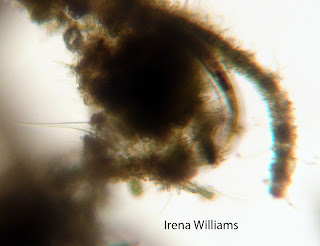
I did my last observation on Wednesday, November 10, 2010.  I didn't notice any changes since the previous week.  There were still quite a few Tachysomas throughout the MicroAquarium, as well as several Seed Shrimp and Cyclopses.  I was finally able to spot a Nematoda, which may be the mysterious organism I kept spotting but not being able to see clearly enough to identify.

Nematoda (identified from Thorp and Covich 1991)

Tachysoma

Citations:
Thorp, James, and Alan Covich. Ecology and Classification of North American Freshwater Invertebrates. 1991.  San Diego, New York, Boston, London, Sydney, Tokyo, Toronto: Academic Press, Inc. p. 250
Thursday, November 4, 2010

Week 3 Observation


This week I didn't plan to take any pictures, but I found some interesting activity in my MicroAquarium when I observed it on Wednesday, November 3.  First, I noticed that there were a lot of dead seed shrimp inside the bladders of the Utricularia giba bladder.  Then, I found that there was a live cyclops trapped inside one of the bladders.  When I first began observing the cyclops, it was moving rather frantically around the bladder, but by the time I started video taping, it had settled down.  In the video, you can see it kick its legs and then be still again.  I was also able to video tape some other cypcloses and some trachyosomas swimming in the area around the bladder.  Unfortunately, my video editing skills are not very good, but if I have a chance later, I will try again to improve the video quality.

Tuesday, November 2, 2010

Week 2 Observation

I observed my MicroAquarium under the microscope on Wednesday, October 27.  I observed several of the same organisms that I had seen the previous week.  Several of the seed shrimp had died and settled near the bottom of the tank.

The previous Friday, one Beta Food Pellet had been inserted into the microaquarium.  The Beta Food Pellet was "Atison's Betta Food" made by Ocean Nutrition, Aqua Pet Americas, 3528 West 500 South, Salt Lake City, UT 84104. Ingredients: Fish meal, wheat flower, soy meal, krill meal, minerals, vitamins and preservatives. Analysis: Crude Protein 36%; Crude fat 4.5%; Crude Fiber 3.5%; Moisture 8% and Ash 15%. (McFarland 2010).

There were quite a few trachyosomas in the area around the Beta Food.  I also observed a few cyclopses in the area.  I think they were probably feeding on the trachyosomas.

cyclops (identified from Rains and Russell 1996)


seed shrimp (identified from Rains and Russell 1996)

Citations:
McFarland Ken. “Botany 111 2010.” http://botany1112010.blogspot.com/.  University of Tennessee/Botany 111. 10/04/2010.

Rains Kenneth G, Russell Bruce J.  1996.  Guide to Microlife.  Franklin Watts.  p. 204, 209.

Sunday, October 24, 2010

Week 1 Observation


I observed my MicroAquarium through a microscope on Wednesday, October 20.  I saw quite a few seed shrimp (pictured above) swimming around the areas near the plants (identified from Rains and Russell 1996).  This is the organism I described last time as looking like a clam with hairs coming out of the space between the two parts of the shell.


I was able to photograph an organism that may be a trachyosoma (pictured below).  Since I didn't get a very clear picture, I wasn't able to positively identify it.


I also observed the "contact lens-like" organisms I mentioned in my last post, but I was unable to locate them long enough to take a picture of them.


In general, I noticed that the areas of the water around the plants and soil were full of life, but the clear water didn't appear to have any living organisms.  It was very interesting to look through the microscope and see the organisms moving around in the water, but extremely difficult to take pictures of them, since they move very quickly and tend to spend most of their time moving among the plants.  Interestingly, I did not see any of the "tick-like" organisms that I spotted when I first set up my MicroAquarium.


Citations:
Rains Kenneth G, Russell Bruce J.  1996.  Guide to Microlife.  Franklin Watts.  p. 204, 209.

Friday, October 15, 2010

MicroAquarium Setup

I set up a MicroAquariumTM on Wednesday, October  13.  I put a glass tank in a stand holder and then took a sample of soil and water that had been collected from the pond at University of Tennessee Hospital at Cherokee Trail.  According to the class website (McFarland), the water was collected from the pond, which has full sun exposure and is located at N35 56.305 W83 56.717 850 ft, on October 10.  Using a pipet, I collected some soil, some water from the middle, and some water from the surface of the larger sample.  I then squeezed the water into the glass tank until it was about 2/3 full.  Next, I selected a sample of a moss, Fontinalis sp., which was collected from the natural spring at Carters Mill Park on October 10 (McFarland) and added the Fontinalis sp. to my MicroAquariumTM.  I then inserted a sample of a flowering plant, Utricularia gibba L., which was grown in tanks outside the greenhouse in Hesler Biology Building after having been taken from the south shore of Spain Lake in White County, TN (McFarland).  I then carefully placed my MicroAquariumTM under the microscope to observe the organisms inside.
I saw several organisms moving around in the water.  The first I saw looked like a contact lens with green bumps on it, and it flipped around in three dimensions and suddenly “swam” out of the field of vision.  Then, I saw something that looked like a flat, green drawing of a tick.  It appeared to have about five “legs” and seemed to move in sudden jerks.  The third organism I saw moving looked somewhat like a clam with “legs” coming out of the middle.  It looked dark brownish-green and appeared to be feeding on other organisms in the tank.

Citations: Ken McFarland. “Botany 111 2010.” http://botany1112010.blogspot.com/.  University of Tennessee/Botany 111. 10/04/2010.